Cryptorchidism in dogs means one or both testes never made it down into the scrotum. The retained testis carries a real risk of developing a tumor later in life. Veterinarians agree the undescended testis should come out. But what about a normal, descended testis sitting in the scrotum? Current research offers reassurance for young dogs and a more cautious picture for older ones, which opens the door to hormone-sparing choices like vasectomy.
What is cryptorchidism in dogs?
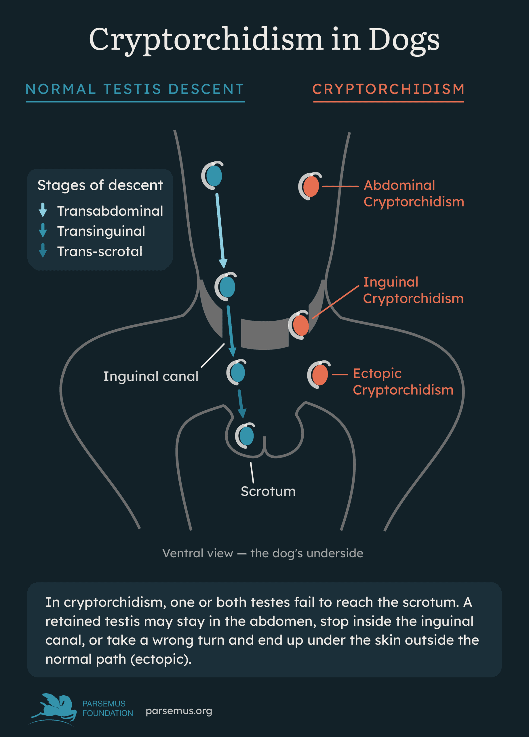
Cryptorchidism refers to a retained testis where one or both testes fail to complete their normal migration into the scrotum. This migration is under hormonal control. The testes descend from the abdomen, through the inguinal canal, and into the scrotum. In a healthy puppy, both testes should be felt in the scrotum by six to eight weeks of age. If a testis is still missing by six months, most veterinarians consider it permanently retained.
A retained testis can end up in one of a few places. It might stay inside the abdomen, sit in the inguinal canal near the groin, or land just outside the canal in an abnormal spot under the skin. All three situations get lumped under the umbrella term cryptorchidism. Inguinal canal placement is the most common type in dogs.
This condition is fairly common. Reported rates range from under 1% to as high as 15% of dogs, depending on the study and breed mix. Cryptorchidism affects purebred dogs and small or toy breeds most frequently. Chihuahuas, toy poodles, Pomeranians, boxers, Pekingese, and Yorkshire terriers all show up repeatedly on the high-risk list. It also frequently affects German Shepherds.
Most cases are unilateral, meaning only one testis fails to descend while the other descends normally. That pattern accounts for roughly 75–81% of cases. Cryptorchidism affects the right testis about twice as often as the left. Researchers also believe it has a hereditary, sex-limited autosomal recessive basis (only affecting males with two copies of the recessive gene). Because cryptorchid dogs can pass the predisposition to their offspring, affected dogs and their siblings are not recommended for breeding.
Courtesy of Dogs4Motion Academy
Why cryptorchidism in dogs is a health concern
The biggest worry with a retained testis is cancer. A cryptorchid testis develops tumors far more often than a normally descended one. Multiple studies report cryptorchid dogs face 9 to nearly 14 times the risk of testicular cancer compared with dogs whose testes descended normally. Testicular tumors are also found at younger ages in cryptorchid dogs. In retained testes, Sertoli cell tumors and seminomas dominate. One 12-year retrospective study found Sertoli cell tumors made up a much larger share of tumors in cryptorchid testes than in the general testicular tumor population.
Why do misplaced testes turn cancerous more often? Researchers have not settled on one answer. Two main ideas compete. The first blames heat: a testis inside the warm abdomen sits several degrees hotter than one in the scrotum. That extra heat may disrupt normal cell maturation over time. The second idea points to a shared developmental problem. Under this theory, the same genetic or environmental disruption that stopped the testis from descending in the first place also primes its cells toward cancer, independent of temperature.
Both mechanisms have supporting evidence, but neither has definitive proof, and no consensus exists yet. This uncertainty doesn’t change the bottom line, though. Removing the retained testis is standard practice because its cancer risk is well documented, even if the cause isn’t.
Why someone might want to preserve the descended testis
Here’s where the question gets more interesting for owners of unilaterally cryptorchid dogs. If the retained testis needs to come out, does a veterinarian need to remove the normal, scrotal testis too?
Traditional veterinary advice says yes: remove both, mainly to guarantee sterility and prevent the dog from passing on the (heritable) condition. But removing a healthy testis also removes its testosterone output. A growing body of research links that loss of natural hormones to real downside risks.
For example, a 2023 survey study by Zink and colleagues compared thousands of dogs across five reproductive categories. They included intact, castrated, spayed, vasectomized, and hysterectomized (ovary-sparing spay) dogs. Dogs with longer lifetime exposure to their own gonadal hormones, whether intact or sterilized by a hormone-sparing method, had lower odds of general health problems and of both problematic and nuisance behaviors than fully gonadectomized dogs. This is the concept behind hormone-sparing sterilization, including vasectomy for male dogs and ovary-sparing spay (hysterectomy) for females. These sterilization methods preserve natural hormones (see our pages on Hormone-Sparing Sterilization).
A large-breed dog like a Golden Retriever, or any breed with documented sensitivity to gonad removal, is exactly the kind of dog whose owner might want to preserve testicular hormone production of the descended testis while removing an undescended testis.
“A vasectomy can be performed”
Dr. Robert McCarthy is a veterinary surgeon with expertise in canine vasectomy and hysterectomy. He’s an Adjunct Clinical Associate Professor at Tufts Cummings School of Veterinary Medicine. He co-authored a 2023 chapter on Cryptorchidism in Small Animal Soft Tissue Surgery. He noted that “Even if the condition is unilateral, both the descended and undescended testicles should be removed to prevent hereditary transmission of this defect. Alternatively, if an owner prefers to preserve the dog’s gonadal hormones, then a vasectomy can be performed on the descended testicle.”
The tough question: is it risky to keep the descended testis?
This brings us to the heart of the matter. Two studies, using two different approaches, help answer it, but they don’t fully agree.
- Veronesi et al. (2009) studied 10 young dogs, aged 1 to 2 years. Each had one retained testis (either abdominal or ectopic) and one normal scrotal testis. The team examined both testes under the microscope and used immunohistochemistry, a lab technique that uses antibody stains to reveal specific proteins inside cells, to check for markers of immaturity or abnormal development. The retained testis showed clear damage: Sertoli cell-only tubules (seminiferous tubules missing their germ cells entirely), reduced size, and reduced firmness. The scrotal testis, however, looked completely normal. They had full sperm production, mature Sertoli cells, and no signs of the immaturity markers seen in the retained testis. The researchers concluded that, at least in young dogs, continued monitoring of the healthy scrotal testis is a reasonable alternative to removing it.
- Pecile et al. (2021) took a wider view. Their study included 26 dogs spanning a much broader age range, from 5 months to 13 years old (median age 3 years), with both unilateral and bilateral cryptorchidism represented. They tested both retained and scrotal testes for specific markers of the fetal/neonatal period linked to cancerous tissue. As expected, the retained testis showed significantly more marker positivity than scrotal ones. But here’s the finding that matters most for this question: some scrotal testes from unilaterally cryptorchid dogs, even ones with no visible anatomical problems, also tested positive for some of these markers. The authors interpreted this as a possible early, subclinical warning sign and recommended considering bilateral removal as a precaution.
Interpreting the studies
So which study is right? Both, in a sense, because they looked at different populations. Veronesi’s dogs were all young, 1 to 2 years old. Pecile’s study included dogs up to 13 years old, and several analyses found marker expression increased with age. It’s possible that a young, unilaterally cryptorchid dog’s scrotal testis really is unaffected, as Veronesi found. Meanwhile, age and time may introduce subtle cellular changes later in life, as Pecile’s broader sample suggests.
Neither study followed dogs over time to see whether marker-positive scrotal testes actually went on to develop tumors. Nor did they compare cancer rates directly between the retained and contralateral scrotal testes within the same dogs. In fact, a comprehensive 2024 review of the topic points out that no study in dogs has directly compared cancer outcomes between a cryptorchid testis and its normal, contralateral scrotal partner.
Cryptorchidism also afflicts humans, and research has confirmed that a normally descended testis in a patient with cryptorchidism does not have an increased risk of cancer. While the human condition does not always translate to canines, extensive research in humans can add to our limited understanding of cryptorchidism in dogs.
Overall, the risk of cancer in the scrotal testis appears low for unilateral cryptorchid dogs, especially in younger individuals, but it may not stay low indefinitely.
Vasectomy: a middle path
This is exactly where a vasectomy can help. A vasectomy cuts or blocks the vas deferens, the tube that carries sperm, without removing the testis or its hormone-producing tissue. For a unilaterally cryptorchid dog, that means a veterinarian can remove the higher-risk retained testis through castration. In the same anesthetic event, they can also perform a vasectomy on the remaining, normal scrotal testis.
The dog becomes fully sterile, so he cannot pass on any genes related to cryptorchidism. The scrotal testis keeps producing testosterone for life, preserving the potential musculoskeletal, metabolic, and behavioral benefits associated with natural hormone exposure. This combined approach directly addresses the documented cancer risk of the retained testis while sidestepping the hormone loss that comes with full castration.
What to watch for if you keep the descended testis
If you and your veterinarian decide to keep the scrotal testis, ongoing monitoring matters. Here’s what to watch for:
- Regular palpation. Check the remaining testis monthly for changes in size, firmness, or shape. A healthy testis should feel smooth, firm, and roughly the same size over time.
- Annual veterinary exams. Ask your vet to palpate the testis at every checkup, especially as your dog ages, since tumor risk in intact male dogs generally rises with age.
- Signs of feminization. Sertoli cell tumors can secrete excess estrogen. Watch for symmetrical hair loss, nipple enlargement, attractiveness to other male dogs, or a pendulous prepuce. These signs warrant a prompt vet visit.
- Enlargement or asymmetry. Have your veterinarian check for any sudden change in the testis size or texture with ultrasound.
- Breed-specific vigilance. If your dog belongs to a breed already prone to testicular tumors, discuss the appropriate monitoring schedule with your veterinarian.
None of these signs are unique to a testis that was once part of a cryptorchid pair. They’re the same signs any owner of an intact male dog should watch for. That said, closer attention is reasonable given the unresolved questions raised by the immunohistochemistry research.
Take-home message on cryptorchidism in dogs
Cryptorchidism is common in dogs, especially in small breeds. The retained testis carries a clear, well-documented cancer risk that justifies its removal. The evidence of cancer for a descended testis is low, particularly in young dogs. However, some subtle changes may appear with age. For owners who want to avoid the retained testis’s risk while still preserving natural hormone production, pairing removal of the undescended testis with a vasectomy of the scrotal testis offers a practical, hormone-sparing path to full sterility. As with any individualized medical decision, talk with a veterinarian familiar with hormone-sparing sterilization about your dog’s breed, age, and health history before deciding. You can find experienced professionals on our Veterinary Directory.
Bibliography
Gradil, C. and McCarthy, R. Cryptorchidism. In Small Animal Soft Tissue Surgery, E. Monnet (Ed.). 2023. https://doi.org/10.1002/9781119693741.ch59
Hayes HM Jr, Pendergrass TW. Canine testicular tumors: epidemiologic features of 410 dogs. Int J Cancer. 1976;18(4):482–487. https://onlinelibrary.wiley.com/doi/10.1002/ijc.2910180413
Liao AT, Chu PY, Yeh LS, Lin CT, Liu CH. A 12-year retrospective study of canine testicular tumors. J Vet Med Sci. 2009;71(7):919–923. https://doi.org/10.1292/jvms.71.919
Pecile A, Groppetti D, Pizzi G, Banco B, Bronzo V, Giudice C, Grieco V. Immunohistochemical insights into a hidden pathology: canine cryptorchidism. Theriogenology. 2021;176:43–53. https://doi.org/10.1016/j.theriogenology.2021.09.011
Romagnoli SE. Canine cryptorchidism. Vet Clin North Am Small Anim Pract. 1991;21(3):533–544. https://doi.org/10.1016/s0195-5616(91)50059-0
Soto-Heras S, Reinacher L, Wang B, Oh JE, Bunnell M, Park CJ, Hess RA, Ko CJ. Cryptorchidism and testicular cancer in the dog: unresolved questions and challenges in translating insights from human studies. Biol Reprod. 2024;111(2):269–291. https://doi.org/10.1093/biolre/ioae075
Veronesi MC, Riccardi E, Rota A, Grieco V. Characteristics of cryptic/ectopic and contralateral scrotal testes in dogs between 1 and 2 years of age. Theriogenology. 2009;72(7):969–977. http://dx.doi.org/10.1016/j.theriogenology.2009.06.016
Yates D, Hayes G, Heffernan M, Beynon R. Incidence of cryptorchidism in dogs and cats. Vet Rec. 2003;152(16):502–504. https://doi.org/10.1136/vr.152.16.502
Zink C, Delgado MM, Stella JL. Vasectomy and ovary-sparing spay in dogs: comparison of health and behavior outcomes with gonadectomized and sexually intact dogs. J Am Vet Med Assoc. 2023;261(3):366–374. https://doi.org/10.2460/javma.22.08.0382